Contaminated or Not? Guidelines for Interpretation of Positive Blood Cultures
Weinstein MP. Contaminated or Not? Guidelines for Interpretation of Positive Blood Cultures. PSNet [internet]. Rockville (MD): Agency for Healthcare Research and Quality, US Department of Health and Human Services. 2008.
Weinstein MP. Contaminated or Not? Guidelines for Interpretation of Positive Blood Cultures. PSNet [internet]. Rockville (MD): Agency for Healthcare Research and Quality, US Department of Health and Human Services. 2008.
The Case
A 62-year-old man with type 2 diabetes mellitus, chronic kidney disease, and a history of ventricular tachycardia with an automated implantable cardiac defibrillator (AICD) came to his primary care physician (PCP) with symptoms of shaking, weakness, and vomiting. He denied fevers. The physical examination was unremarkable except for the presence of chronic peripheral neuropathy. The physician ordered routine blood tests and 2 peripheral blood cultures, diagnosed the patient with a nonspecific viral syndrome, and sent him home.
The routine laboratory tests done that day revealed only a normocytic anemia. However, 5 days later, the PCP was notified that both sets of blood cultures were growing Corynebacterium spp. Uncertain of how to interpret the result (as this bacteria may represent contaminated blood cultures rather than a true cause of disease), the PCP contacted an infectious disease specialist, who recommended hospitalization. The patient was hospitalized, seen by a different infectious disease specialist, and started on IV antibiotics. The patient's subsequent evaluation revealed no evidence of infection, including an unremarkable abdominal CT scan and a normal transthoracic echocardiogram (TTE). Repeat blood cultures (drawn before antibiotics were begun) remained negative. The patient was clinically stable, so the antibiotics were stopped and the patient was discharged to home. The physicians assumed that the Corynebacterium was a contaminant from the skin.
One month later, the patient presented to the emergency department (ED) with nausea and vomiting. His physical examination and laboratory test results were unremarkable. His symptoms improved with IV fluids, and he was discharged after an 18-hour stay.
Two days later, 2 out of 2 blood cultures drawn at that ED visit started growing Corynebacterium spp. That evening, the results were reported to a covering physician who was unfamiliar with the patient or previous culture results. The physician assumed that the blood cultures were contaminated from the skin and took no action.
Three weeks later, the patient was readmitted after being shocked by his defibrillator (AICD). A transesophageal echocardiogram (TEE) revealed a tricuspid vegetation and blood cultures again showed Corynebacterium spp. (the final speciation was never determined). Diagnosed with subacute bacterial endocarditis and treated with IV vancomycin, the patient made a full recovery.
The Commentary
The case history that forms the basis for this commentary illustrates several of the important complexities and inefficiencies of modern medicine, some of which resulted in medical errors. These include:
- A patient with multiple underlying medical problems that predispose to infection;
- Isolation of a microorganism from blood cultures that in most circumstances would represent contamination but, in this instance, represented a clinically important pathogen that caused a potentially life-threatening infection;
- Misinterpretation of the clinical significance of the positive blood culture result;
- Failure of the primary and covering physicians to communicate effectively, ultimately resulting in delayed diagnosis and increased patient morbidity.
Although each needs to be appropriately addressed to prevent similar errors, this commentary will focus primarily on the interpretation and potential misinterpretation of positive blood cultures.
Blood Culture Contamination
Physicians and clinical microbiologists have long appreciated that blood cultures are perhaps the most important laboratory tests to diagnose serious infections. In recent years, it has also become apparent that contaminated (i.e., the presence of a pathogen from outside the blood stream) blood cultures are common, leading to falsely positive test results.(1,2) Contaminated blood cultures constitute as many as half or more of all positive blood cultures in some centers, are very costly to patients and the health care system,(3) and are confusing for clinicians.(4,5)
There are numerous reasons why blood cultures are contaminated so frequently. Perhaps the most important factor is the failure of the health care worker (HCW) to use strict aseptic technique when obtaining the blood specimen. Studies have shown that trained phlebotomists or blood culture teams have fewer contaminated blood cultures than other HCWs.(5-7) A second factor is the antiseptic agent itself; tincture of iodine and chlorhexidine gluconate are more effective at skin sterilization than iodophor (povidone iodine) preparations.(8-11) A third factor is the means by which blood is obtained for culture. In recent years, there has been a trend toward obtaining blood cultures from existing indwelling intravenous catheters or other access devices (e.g., ports). However, blood cultures obtained in this fashion are contaminated more frequently than those obtained by peripheral venipuncture.(12-14) Fourth, modern blood culture systems and media that incorporate antibiotic-binding resins or activated charcoal, while detecting more true pathogens, also have been shown to greatly enhance the detection of coagulase-negative staphylococci, the most common blood culture contaminants.(5) Finally, blood culture techniques changed after recognition that HIV is a blood-borne pathogen. In the pre-HIV era, the needle used to obtain the blood culture was removed and a second sterile needle was placed on the syringe for inoculation of the blood culture bottles. To reduce the risk of needlestick injury associated with changing needles, the standard culture method now employs a single needle that is used for obtaining blood and inoculating the culture vial. Although several studies initially showed that the single needle technique was not associated with increased contamination rates, a subsequent meta-analysis showed a contamination rate of 3.7% with the 1-needle method versus 2.0% with the 2-needle technique.(15)
Guidelines for Interpretation of Positive Blood Cultures
Some clinical and laboratory tools can aid physicians and microbiologists in deciding whether a blood isolate is a pathogen or a contaminant. Obviously, the presence of predisposing factors and a consistent clinical presentation can help clinicians interpret test results. The identity of the microorganism also provides important information (Table), and a predictive model has confirmed this.(16) Microorganisms that always or nearly always (greater than or equal to 90%) represent true infection when isolated from blood cultures include S. aureus, S. pyogenes, S. agalactiae, S. pneumoniae, E. coli and other members of the family Enterobacteriaceae, P. aeruginosa, B. fragilis group, and Candida species. In contrast, coagulase-negative staphylococci (CoNS), Corynebacterium species, Bacillus species other than anthracis, and P. acnes usually represent contamination. Isolation of the latter microorganisms, mostly commonly with CoNS but also with corynebacteria (as in the case presented here), may confuse clinicians. Corynebacterium species are part of the normal human skin flora, so they typically do not cause true invasive disease. But Corynebacterium can cause clinically significant infections in the presence of medical devices such as joint prostheses, catheters, ports, vascular grafts, prosthetic heart valves, pacemakers, and AICDs (as in this case).
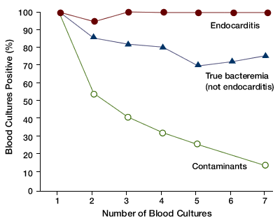
The number of blood culture sets that grow a particular microorganism, especially when measured as a function of the total number of blood cultures obtained, has proved to be a very useful aid in interpreting the clinical significance of positive blood cultures (Figure).(2,17,18) In true endovascular (within the blood vessels) infections and other blood stream infections (BSIs), either all or most of the blood cultures obtained at the time of diagnosis will be positive, whereas when a blood culture is contaminated, usually only one of several blood culture sets will be positive. As the Figure illustrates and this statement implies, this diagnostic maxim has no utility if only a single blood culture is obtained. The value of multiple cultures largely flows from probability considerations: Most institutions have contamination rates in the range of 3% per blood culture drawn. It follows, then, that the probability of recovering the same microorganism in 2 culture sets from a patient, and of that organism being a contaminant, is less than 1 in 1000 (0.03 x 0.03 = 0.0009). The clinician can be quite confident, then, that 2 out of 2 blood cultures positive with the same pathogen, even one that is commonly a contaminant, represents real disease, assuming that the 2 blood cultures were obtained from separate venipunctures or catheter draws.
Reducing Contamination
We cannot eliminate blood culture contamination entirely, but it is possible for institutions to reduce contamination rates. One step is to use more efficacious antiseptic preparations. Povidone iodine preparations (iodophors) require 1.5 to 2 minutes of contact time to produce maximum antiseptic effect, whereas iodine tincture and chlorhexidine gluconate only require 30 seconds.(5,19) Many HCWs who obtain blood cultures are in a hurry, do not understand the importance of antiseptic contact time, and are unlikely to wait up to 2 minutes before obtaining blood for culture. Although the evidence-base has limitations,(20) the Clinical and Laboratory Standards Institute, a consensus organization that publishes guidelines based on best available data, recommends tincture of iodine, chlorine peroxide, and chlorhexidine gluconate over povidone-iodine and further states that iodine tincture and chlorhexidine gluconate are probably equivalent.(11) Malani and colleagues (20) identified a possible benefit related to the use of commercially marketed prepackaged skin antiseptic kits. However, available data are limited, and I believe that no firm recommendations regarding these prepackaged kits can be made at this time.
Hospitals may also be able to reduce blood culture contamination rates by utilizing trained phlebotomists or blood culture teams to obtain blood for culture rather than using random nursing personnel, nondegree nursing assistants, medical students, and resident physicians to obtain these specimens.(5-7,21) Laboratory-trained phlebotomists and blood culture teams can be better trained and focused on correct antiseptic technique. Additionally, their individual contamination rates can be monitored as part of an institution's performance improvement program.
Because approximately half of all positive blood cultures in most institutions represent contamination, laboratories should develop policies and procedures to limit the evaluation of likely contaminants.(1,5,22) For example, if only a single blood culture grows a coagulase-negative staphylococcus, Bacillus spp., Corynebacterium spp., Propionibacterium spp., viridans group streptococcus, Micrococcus spp., or Aerococcus spp., the likelihood of contamination is high, and full identification of the microorganism as well as susceptibility testing should not be done unless there is direct communication between the physician caring for the patient and the laboratory director.(5,11,22)
Regarding the case history presented herein, a few issues are worth emphasizing. Microorganisms that are most often contaminants can, in the right clinical setting, be clinically significant pathogens. The initial management of this patient—deeming the initial positive blood cultures to be significant—was reasonable in my judgment. When both the imaging studies and repeat blood cultures prior to antibiotics were negative, treatment was stopped and the patient was observed. However, 1 month later, the patient again had 2 of 2 blood cultures positive for Corynebacterium spp. No action was taken by the covering physician, even though the probability of contamination was less than 1 in 1000. I believe that this represented an interpretation error. Apparently, the PCP was not made aware of this event (a communication error), and no medical intervention occurred, leading to delayed diagnosis and treatment of the patient. Fortunately, the patient suffered no permanent harm, but patient morbidity and cost to the health care system could have been prevented had these errors not occurred.
Take-Home Points
- Blood culture contamination is common, constituting up to half of all positive blood cultures at some institutions.
- The identity of the organism isolated can help in determining if the culture is contaminated, as some organisms rarely cause BSIs.
- The number of blood cultures that yield a particular organism can help predict true infections. For example, if 2 sets of blood cultures obtained by separate venipunctures in the same time frame are positive with the same organism, the probability of contamination is less than 1 in 1000.
- Institutions can reduce blood culture contamination by using the most effective antiseptic agents and utilizing dedicated personal to draw blood cultures.
Melvin P. Weinstein, MD Professor of Medicine and Pathology Robert Wood Johnson Medical School University of Medicine and Dentistry of New Jersey
References
1. Richter SS, Beekmann SE, Croco JL, et al. Minimizing the workup of blood culture contaminants: implementation and evaluation of a laboratory-based algorithm. J Clin Microbiol. 2002;40:2437-2444. [go to PubMed]
2. Weinstein MP, Towns ML, Quartey SM, et al. The clinical significance of positive blood cultures in the 1990s: a prospective comprehensive evaluation of the microbiology, epidemiology, and outcome of bacteremia and fungemia in adults. Clin Infect Dis. 1997;24:584-602. [go to PubMed]
3. Bates DW, Goldman L, Lee TH. Contaminant blood cultures and resource utilization. The true consequences of false-positive results. JAMA. 1991;265:365-369. [go to PubMed]
4. Rupp ME, Archer GL. Coagulase-negative staphylococci: pathogens associated with medical progress. Clin Infect Dis. 1994;19:231-243. [go to PubMed]
5. Weinstein MP. Blood culture contamination: persisting problems and partial progress. J Clin Microbiol. 2003;41:2275-2278. [go to PubMed]
6. Weinbaum FI, Lavie S, Danek M, Sixsmith D, Heinrich GF, Mills SS. Doing it right the first time: quality improvement and the contaminant blood culture. J Clin Microbiol. 1997;35:563-565. [go to PubMed]
7. Surdulescu S, Utamsingh D, Shekar R. Phlebotomy teams reduce blood-culture contamination rate and save money. Clin Perform Qual Health Care. 1998;6:60-62. [go to PubMed]
8. Little JR, Murray PR, Traynor PS, Spitznagel E. A randomized trial of povidone-iodine compared with iodine tincture for venipuncture site disinfection: effects on rates of blood culture contamination. Am J Med. 1999;107:119-125. [go to PubMed]
9. Mimoz O, Karim A, Mercat A, et al. Chlorhexidine compared with povidone-iodine as skin preparation before blood culture: a randomized controlled trial. Ann Intern Med. 1999;131:834-837. [go to PubMed]
10. Strand CL, Wajsbort RR, Sturmann K. Effect of iodophor vs. iodine tincture skin preparation on blood culture contamination rate. JAMA. 1993;269:1004-1006. [go to PubMed]
11. Wilson ML, Mitchell M, Morris AJ, et al. Principles and Procedures for Blood Cultures; Approved Guideline. Wayne, PA: Clinical and Laboratory Standards Institute; 2007. ISBN: 1562386417.
12. Bryant JK, Strand CL. Reliability of blood cultures collected from intravascular catheter versus venipuncture. Am J Clin Pathol. 1987;88:113-116. [go to PubMed]
13. DesJardin JA, Falagas MA, Ruthazer R, et al. Clinical utility of blood cultures drawn from indwelling central venous catheters in hospitalized patients with cancer. Ann Intern Med. 1999;131:641-647. [go to PubMed]
14. Everts RJ, Vinson EN, Adholla PO, Reller LB. Contamination of catheter-drawn blood cultures. J Clin Microbiol. 2001;39:3393-3394. [go to PubMed]
15. Spitalnic SJ, Woolard RH, Mermel LA. The significance of changing needles when inoculating blood cultures: a meta-analysis. Clin Infect Dis. 1995;21:1003-1006. [go to PubMed]
16. Bates DW, Lee TH. Rapid classification of positive blood cultures. Prospective validation of a multivariate algorithm. JAMA. 1992;267:1962-1966. [go to PubMed]
17. MacGregor RR, Beaty HN. Evaluation of positive blood cultures. Guidelines for early differentiation of contaminated from valid positive cultures. Arch Intern Med. 1972;130:84-87. [go to PubMed]
18. Weinstein MP, Reller LB, Murphy JR, Lichtenstein KA. The clinical significance of positive blood cultures: a comprehensive analysis of 500 episodes of bacteremia and fungemia in adults. I. Laboratory and epidemiologic observations. Rev Infect Dis. 1983;5:35-53. [go to PubMed]
19. King TC, Price PB. An evaluation of iodophors as skin antiseptics. Surg Gynecol Obstet. 1963;116:361-365. [go to PubMed]
20. Malani A, Trimble K, Parekh V, Chenoweth C, Kaufman S, Saint S. Review of clinical trials of skin antiseptic agents used to reduce blood culture contamination. Infect Control Hosp Epidemiol. 2007;28:892-895. [go to PubMed]
21. Schifman RB, Pindur A. The effect of skin disinfection materials on reducing blood culture contamination. Am J Clin Pathol. 1993;99:536-538. [go to PubMed]
22. Baron EJ, ed. Cumitech 1C: Blood Cultures IV. Washington, DC: ASM Press; 2005.
Table
Table. Microorganisms Isolated from Blood Categorized According to Clinical Significance.
(Adapted with permission. Original table © 1997 by the University of Chicago. [Weinstein MP, Towns ML, Quartey SM, et al. The clinical significance of positive blood cultures in the 1990s: a prospective comprehensive evaluation of the microbiology, epidemiology, and outcome of bacteremia and fungemia in adults. Clin Infect Dis. 1997;24:584-602.])
| Microorganism | No. (%) of Isolates per Indicated Category | ||
|---|---|---|---|
| (No. of Isolates) | True Pathogen | Contaminent | Unknown |
| Staphylococcus aureus (204) | 178 (87.2) | 13 (6.4) | 13 (6.4) |
| Coagulase-negative staphylococcus (703) | 87 (12.4) | 575 (81.9) | 41 (5.8) |
| Streptococcus pneumoniae (34) | 34 (100) | 0 | 0 |
| Viridans streptococci (71) | 27 (38.0) | 35 (49.3) | 9 (12.7) |
| Other streptococci (31) | 21 (67.7) | 6 (19.4) | 4 (12.9) |
| Enterococcus spp. (93) | 65 (69.9) | 15 (16.1) | 13 (14.0) |
| Corynebacterium spp. (53) | 1 (1.9) | 51 (96.2) | 1 (1.9) |
| Bacillus spp. (12) | 1 (8.3) | 11 (91.7) | 0 |
| Escherichia coli (143) | 142 (99.3) | 0 | 1 (0.7) |
| Klebsiella pneumoniae (65) | 65 (100) | 0 | 0 |
| Other enteric gram-negative bacteria (108) | 104 (96.3) | 1 (0.9) | 3 (2.8) |
| Pseudomonas aeruginosa (55) | 53 (96.4) | 1 (1.8) | 1 (1.8) |
| Propionibacterium acnes (48) | 0 | 48 (100) | 0 |
| Other Gram-positive anaerobes including Clostridium spp. (35) | 19 (54.3) | 15 (42.8) | 1 (2.9) |
| Bacteroides fragilis group (18) | 16 (88.9) | 0 | 2 (11.1) |
| Other Gram-negative anaerobes (5) | 2 (40) | 2 (40) | 1 (20) |
| Candida spp. (60) | 56 (93.3) | 0 | 4 (6.7) |
| Cryptococcus neoformans (8) | 8 (100) | 0 | 0 |
Figure
Figure. Patterns of positivity in sequential blood cultures as an aid to the differentiation of clinically important infection versus contamination. (Reprinted with permission. Figure © 1983 by the University of Chicago. [Weinstein MP, Reller LB, Murphy JR, Lichtenstein KA. The clinical significance of positive blood cultures: a comprehensive analysis of 500 episodes of bacteremia and fungemia in adults. I. Laboratory and epidemiologic observations. Rev Infect Dis. 1983;5:35-53.])


