Abdominal Pain in Early Pregnancy
Kilpatrick CC. Abdominal Pain in Early Pregnancy. PSNet [internet]. Rockville (MD): Agency for Healthcare Research and Quality, US Department of Health and Human Services. 2015.
Kilpatrick CC. Abdominal Pain in Early Pregnancy. PSNet [internet]. Rockville (MD): Agency for Healthcare Research and Quality, US Department of Health and Human Services. 2015.
Case Objectives
- Recognize when nausea and vomiting in pregnancy is abnormal.
- Identify the most common causes of non-obstetric abdominal pain and acute abdomen in early pregnancy.
- Review the diagnosis of appendicitis in pregnancy.
- Discuss a diagnostic imaging algorithm for pregnant women with suspected appendicitis.
Case & Commentary—Part 1
A 34-year-old woman who was 14 weeks pregnant presented to the emergency department (ED) with 5 days of nonspecific abdominal pain, nausea, and vomiting. On examination, she appeared well with normal vital signs and had some mild diffuse abdominal tenderness. Her white blood cell count was 19,000 cells/μL, and a urinalysis was positive for nitrates and leukocyte esterase (indicating possible infection). She was diagnosed with a urinary tract infection and was discharged on antibiotic therapy. No imaging was performed at this initial visit.
The patient returned the following day with unchanged abdominal pain and more nausea and vomiting. A fetal ultrasound was performed and found normal fetal heart activity. No further testing was done, and she was discharged home with instructions to continue the antibiotics.
Abdominal pain remains the most common reason for emergency department (ED) visits, comprising more than 11% of all visits in 2008.(1) In 2011, 54% of patients that presented to the ED were female, more than 25% were of childbearing age, and the pregnancy rate in the United States is approximately 10% at any given time.(2,3) For these reasons, clinicians that evaluate patients with abdominal pain in the ED should be familiar with common causes of abdominal pain in pregnant women and appreciate when nausea and vomiting in pregnancy is abnormal.
Nausea, vomiting, and abdominal pain are very common in pregnancy. Up to 80% of pregnant women experience nausea and vomiting, most commonly in the first trimester. Symptoms and signs that may indicate another cause include nausea and vomiting persisting past mid-pregnancy (approximately 20 weeks) and associated abdominal pain, fever, or diarrhea. In these instances, a more thorough evaluation is indicated.(4) Due to the enlarging uterus and fetal position/movement, abdominal pain is also common in pregnancy. Warning signs include pain that is localized, abrupt, constant, or severe, or pain that is associated with nausea and vomiting, vaginal bleeding, or fever. With any of these, further investigation into nonpregnancy-related causes is warranted. If any of the warning signs above is present, consultation with an obstetric specialist is recommended.
Women of childbearing age who present to the ED with abdominal pain at minimum should receive a urine pregnancy test, and the location and gestational age of the pregnancy should be determined with ultrasound. Miscarriage and ectopic pregnancy are the most frequent causes of abdominal pain in early pregnancy and are often accompanied by vaginal bleeding.(5,6) Once an early gestational age and intrauterine location is confirmed and miscarriage is ruled out, nonobstetric causes of abdominal pain should be explored, especially if any of the warning signs above are present.
With the exception of ovarian torsion, which is more common in the first trimester, the cause and incidence of non-obstetric abdominal pain in pregnancy varies little by gestational age of the fetus. The following are approximate incidences of some causes of acute abdomen in pregnancy: appendicitis (1/1500 pregnancies), cholecystitis, nephrolithiasis, pancreatitis, and small bowel obstruction (each occur in approximately 1/3000), with ovarian pathology (torsion or symptomatic masses) and uterine leiomyomas less common.(7)
After a history, physical examination, and the pregnancy test and ultrasound, laboratory tests that can assist in narrowing the differential diagnosis—including a complete blood count, liver and pancreatic enzymes, and urinalysis—should be reviewed. The white blood cell count increases to 10,000–14,000 cells/μL in normal pregnancy (and as high as 30,000 cells/μL in labor). However, a left shift in the differential and the presence of bands are abnormal and require further investigation.(8) If clinical signs and symptoms accompanied by laboratory data are not conclusive, prompt imaging may be necessary.
Imaging in pregnancy should begin with ultrasound or magnetic resonance imaging (MRI) as they have no ionizing radiation and have not been linked with fetal harm. Compression ultrasound may be useful in the evaluation of suspected appendicitis, cholecystitis, nephrolithiasis, and ovarian pathology in this setting. However, compression ultrasound becomes less sensitive and specific in pregnancy and relies heavily on the skill of the technician or radiologist. If the ultrasound is nondiagnostic, MRI can be considered as its lack of ionizing radiation also makes it safe for the fetus. MRI can aid in diagnosing acute appendicitis, cholecystitis, bowel obstruction, and ovarian pathology. If MRI is unavailable and there is serious concern for a nonpregnancy-related cause for the abdominal pain, computed tomography (CT) scanning can be performed.
If diagnostic tests that contain ionizing radiation (e.g., CT scanning) are deemed to be clinically necessary, they should not be withheld in the pregnant patient even with the concerns for an increased risk of fetal harm. Although a fetus can be harmed by radiation (including miscarriage, fetal anomalies, fetal growth restriction, intellectual disability, and future childhood cancer), the risk is low, especially at lower radiation doses. During the first 2 weeks of pregnancy, ionizing radiation is associated with an all-or-none type effect (miscarriage or intact survival) based on the radiation dose. After this time period, a dose less than 5 rads is recommended in order to decrease the chances of fetal harm.(9) A normal CT scan of the abdomen and pelvis delivers approximately 1 rad of radiation. As a rule, the least amount of ionizing radiation in necessary diagnostic tests should be utilized in the pregnant patient, and consultation with a radiologist and obstetrician is often helpful to achieve this goal.(9) A full discussion of the fetal risks associated with CT scanning is beyond the scope of this commentary, but a CT scan in this setting should only be obtained after obstetrical consultation.
In this case, at the first visit to the ED, the combination of abdominal pain, nausea, and vomiting appropriately raised concern for a nonpregnancy-related cause and triggered further investigations. The patient was found to have a leukocytosis and a positive urinalysis and was treated for urinary tract infection (UTI). At that first visit, she also should have had a urine pregnancy test and an ultrasound to establish the location and gestational age of the pregnancy. In addition, it would have been reasonable for the providers to have considered imaging, since the symptom constellation—5 days of constant abdominal pain, nausea, vomiting, abdominal tenderness on exam, and an elevated white blood cell count—were incompletely explained by a simple UTI. An ultrasound was performed when the patient returned to the ED and indicated a normal viable pregnancy. However, no further imaging was pursued. Given the severity and persistence of her symptoms despite treatment, a complete abdominal ultrasound (looking for nonpregnancy-related intra-abdominal pathology) would have been appropriate.
Case & Commentary—Part 2
The patient again returned to the ED within 24 hours with persistent complaints. She now appeared more ill, with increased abdominal pain on examination. Magnetic resonance imaging of the abdomen was performed and revealed a ruptured appendix with signs of peritoneal inflammation. The patient was taken immediately to the operating room and was found to have diffuse peritonitis secondary to the ruptured appendix. An emergent laparoscopic appendectomy was performed, which the patient tolerated well. Unfortunately, 3 hours after the operation, she had a spontaneous abortion and subsequent severe bleeding requiring multiple transfusions. She was discharged home days later.
In retrospect, it may not have been appropriate to discharge this patient from the ED after her initial presentation without further evaluation, and she experienced a tragic adverse event.
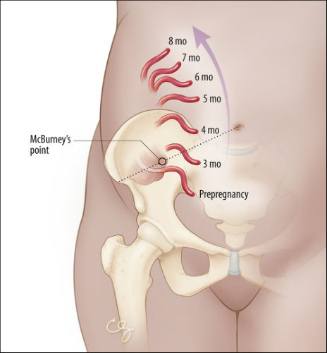
In early pregnancy, the symptoms of appendicitis are identical to those seen in the nonpregnant state: periumbilical pain that migrates to the right lower quadrant, accompanied by anorexia, fever, nausea, and vomiting. With advancing gestational age and in parous women, the diagnosis may become more challenging, largely due to the laxity of the anterior abdominal wall.(10) As the uterus enlarges and fills the peritoneal cavity, a possible cephalad movement of the appendix into the upper quadrant (Figure) can confuse the clinical presentation. Patients may actually present with right upper quadrant pain. Moreover, as the distance between the peritoneum and the appendix increases, peritoneal signs may be masked. Lastly, accurate imaging of the appendix becomes more challenging. Uterine contractions, more common in later gestation, also decrease the ability to accurately elicit peritoneal signs and abdominal tenderness on physical examination. Notwithstanding these variations, clinicians should be aware that right lower quadrant pain remains the most common symptom of appendicitis in pregnant women.(10) Given that appendicitis is the most common cause of acute abdomen in pregnancy, a high index of suspicion is warranted to decrease maternal and fetal morbidity and mortality. In this case, the patient had a somewhat uncommon presentation, with no localization of the abdominal pain. However, the nausea and vomiting and persistent symptoms should have led the clinicians to consider the diagnosis of appendicitis early in the evaluation.
If appendicitis is suspected, diagnostic imaging can help rule in or rule out the diagnosis. Ultrasound is the test of choice in this setting—the diagnosis of appendicitis is made when a noncompressible, blind-ended tubular structure greater than 6 mm is visualized in the right lower quadrant. Unfortunately, the appendix cannot always be visualized with ultrasound, and ultrasound may be less accurate in the setting of a ruptured appendix or with advancing gestational age.(11) The American College of Radiology suggests MRI as the second modality when evaluating for appendicitis in the pregnant patient. The diagnosis is made with visualization of a fluid filled appendix greater than 7 mm in diameter. MRI has high sensitivity and specificity for diagnosing appendicitis.(12) However, MRI technology and expertise are not always available, especially in smaller hospitals or off hours. When this is the case, CT with or without contrast should be used. CT is readily available in most centers, and it has high sensitivity and specificity for the diagnosis of appendicitis. The diagnosis is made by visualization of a fecalith, an enlarged nonfilling tubular structure, or right lower quadrant inflammation. The use of contrast typically enhances diagnostic accuracy with the potential increase in fetal harm (see above). The decision to use contrast should balance the potential fetal harm with the possible delay in diagnosis from nonvisualization of the appendix if contrast is not used. A delay can lead to rupture of the appendix and increase fetal mortality. The fetal loss rate increases from 3%–5% in nonruptured appendicitis to as high as 25% if the appendix ruptures.(13) Maternal morbidity is also higher in the setting of a ruptured appendix.
Institutions should develop an algorithm for optimal imaging of pregnant patients with an acute abdominal process, and this algorithm should be communicated to all the clinicians that care for pregnant patients. The algorithm should take into account best practices as well as the expertise and capability of the radiology team. The goal should be to minimize ionizing radiation and fetal harm while maintaining diagnostic accuracy. The use of the algorithm as well as the clinical outcomes of pregnant patients with abdominal pain should be audited and reviewed. The results of these reviews should be provided to clinicians and could easily be incorporated into institutional meetings that focus on hospital improvement.
The implementation of a checklist may also help to decrease morbidity in the pregnant patient who presents with abdominal pain without introducing any negative effect on safety. Checklists in medicine have been associated with reduced morbidity and mortality, enhanced communication, decreased adverse events, and improved adherence to hospital operating procedures.(14) A computerized checklist could review atypical signs and symptoms in the pregnant patient and prompt clinicians to consider other diagnoses. In this case for example, 5 days of abdominal pain, nausea, and vomiting, with abdominal tenderness on physical examination should have prompted an exploration of diagnoses other than a UTI and may have led to abdominal imaging.
In the above case, in the initial visit, pregnancy location and gestational age should have been documented, and a further investigation into the patient's signs and symptoms should have prompted imaging of the abdomen and pelvis and consultation with an obstetric specialist. When the patient returned with worsening symptoms, appropriate imaging was pursued and revealed acute appendicitis. The error (delay in diagnosis) led to a catastrophic adverse event—the loss of the fetus. A checklist highlighting the warning signs above or an algorithm for early and safe imaging may have led to an earlier diagnosis and a better outcome.
Take-Home Points
- Nausea and vomiting are common in early pregnancy, but symptoms that persist after 16–20 weeks or are accompanied by abdominal pain should be considered abnormal and evaluated.
- Initial imaging in the evaluation of abdominal pain in the pregnant patient should begin with ultrasound or MRI, but ionizing imaging that is clearly indicated by the clinical situation should not be delayed or withheld in the pregnant patient due to concerns of fetal harm.
- Emergency department physicians, in conjunction with obstetric specialists, should develop a diagnostic imaging algorithm for pregnant patients with abdominal pain based on the availability and expertise of their radiologic staff.
- Consultation with an obstetric specialist should be strongly considered in patients with abdominal pain associated with nausea and vomiting in pregnancy.
- Appendicitis remains the most common cause of acute abdomen in pregnancy, and a delay in diagnosis increases fetal mortality.
Charlie C. Kilpatrick, MD Associate Professor of Obstetrics and Gynecology Residency Program Director Baylor College of Medicine, Houston, Texas
Faculty Disclosure: Dr. Kilpatrick has declared that neither he, nor any immediate member of his family, has a financial arrangement or other relationship with the manufacturers of any commercial products discussed in this continuing medical education activity. In addition, the commentary does not include information regarding investigational or off-label use of pharmaceutical products or medical devices.
References
1. Emergency Department Visits for Chest Pain and Abdominal Pain: United States, 1999–2008. Atlanta, GA: National Center for Health Statistics, Centers for Disease Control and Prevention, US Department of Health and Human Services; September 2010. NCHS Data Brief No. 43. [Available at]
2. The National Hospital Ambulatory Medical Care Survey: 2011 Emergency Department Summary Tables. Atlanta, GA: Centers for Disease Control and Prevention, National Center for Health Statistics, Centers for Disease Control and Prevention, US Department of Health and Human Services; November 2014. [Available at]
3. Pregnancy Rates for U.S. Women Continue to Drop. Atlanta, GA: National Center for Health Statistics, Centers for Disease Control and Prevention, US Department of Health and Human Services; December 2013. NCHS Data Brief No. 136. [Available at]
4. McCarthy FP, Lutomski JE, Greene RA. Hyperemesis gravidarum: current perspectives. Int J Womens Health. 2014;6:719-725. [go to PubMed]
5. Doubilet PM, Benson CB, Bourne T, Blaivas M; Society of Radiologists in Ultrasound Multispecialty Panel on Early First Trimester Diagnosis of Miscarriage and Exclusion of a Viable Intrauterine Pregnancy. Diagnostic criteria for nonviable pregnancy early in the first trimester. N Engl J Med. 2013;369:1443-1451. [go to PubMed]
6. Lipscomb GH. Ectopic pregnancy: still cause for concern. Obstet Gynecol. 2010;115:487-488. [go to PubMed]
7. Kilpatrick CC, Monga M. Approach to the acute abdomen in pregnancy. Obstet Gynecol Clin North Am. 2007;34:389-402. [go to PubMed]
8. Kuvin SF, Brecher G. Differential neutrophil counts in pregnancy. N Engl J Med. 1962;266:877-878. [go to PubMed]
9. ACOG Committee on Obstetric Practice. ACOG Committee Opinion #299 (replaces #158, September 1995): guidelines for diagnostic imaging during pregnancy. Obstet Gynecol. 2004;104:647-651. [go to PubMed]
10. Mourad J, Elliot JP, Erikson L, Lisboa L. Appendicitis in pregnancy: new information that contradicts long-held clinical beliefs. Am J Obstet Gynecol. 2000;182:1027-1029. [go to PubMed]
11. Lim HK, Bae SH, Seo GS. Diagnosis of acute appendicitis in pregnant women: value of sonography. AJR Am J Roentgenol. 1992;159:539-542. [go to PubMed]
12. Andreotti RF, Lee SI, Dejesus ASO, et al. ACR Appropriateness Criteria acute pelvic pain in the reproductive age group. Ultrasound Q. 2011;27:205-210. [go to PubMed]
13. Doberneck RC. Appendectomy during pregnancy. Am Surg. 1985;51:265-268. [go to PubMed]
14. Thomassen Ø, Storesund A, Søfteland E, Brattebø G. The effects of safety checklists in medicine: a systematic review. Acta Anaesthesiol Scand. 2014;58:5-18. [go to PubMed]
Figure
Figure. Illustration Showing How Position of Appendix Changes in Pregnancy. (Illustration © 2015 Chris Gralapp.)


